结晶样视网膜色素变性是一个经常被误诊的视网膜疾病
BCD误诊和被低估
误诊(因此患者人数被低估)是许多BCD患者遇到的问题。
研究人员认为,结晶样视网膜色素变性(BCD)的症状与
其他逐渐损害视网膜的眼部疾病相似,因此可能未得到充分
诊断。 Mataftsi et al 2004 报告最初只有六分之一的BCD
患者被正确诊断。
BCD正确诊断率仅为
左右
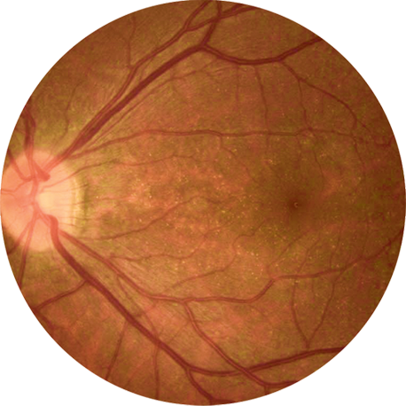
结晶样视网膜色素变性(BCD)有时被眼科医生称为“闪闪发光的视网膜”,原因是BCD患者视网膜中(以及在某些患者的角膜中)存在黄色或黄白色的能够反光晶体和/或复杂的脂质沉积物 。1 尽管视网膜晶体是BCD的特征之一,但它不能用作诊断BCD的唯一临床特征。 首先,有些其他疾病也具有视网膜晶体。 其次,并非所有的BCD患者都有这种临床表现。 此外,BCD与其他一些视网膜疾病具有相似的临床表型,可能被误诊为: 2 3 4 5

1.
视网膜色素变性(RP)
结晶样视网膜色素变性(BCD)是视网膜色素变性(RP)的一个类型,也经常被一般性地诊断为视网膜色素变性(RP)。结晶样视网膜色素变性(BCD)的视野检查,电生理研究和视网膜色素上皮(RPE)/脉络膜变性的临床症状, 与其他类型的视网膜变性类似,属于视网膜色素变性(RP)和相关疾病。此外,视网膜中的晶体沉积可能不明显,尤其是在早期和晚期BCD患者中。 因此,即使是有诊断BCD经验的医生有时也可能会误诊BCD。

2.
无脉络膜症 (Choroideremia)
BCD和无脉络膜症都有进行性脉络膜视网膜变性的症状。 6
3.
其他在视网膜中也有反光晶体样沉积物的视网膜疾病,例如:
- Stargardt Disease (SD) group 3
- dominant forms of RPE65
- RDS – PRPH2 form of retinitis pigmentosa
- 其他晶状体视网膜病变。 视网膜中的晶体沉积不是BCD独有的,并且可能与以下因素有关:
- Primary hyperoxaluria type 1 and type 2
- Cystinosis, particularly the more benign adolescent presentation
- Sjögren-Larsson syndrome
- Infectious Crystalline Keratopathy
- 一些药物也可能造成视网膜上形成晶体(tamoxifen, the anesthetic methoxyflurane,
the oral tanning agent canthaxanthine) - 药物滥用(talc retinopathy)
4.
一些BCD患者报告说,医生告知他们根本没有眼病,并且视网膜中的晶体或异常外观是由于:
- 污染,或
- 不健康的饮食
关于如何改善BCD正确诊断的建议
诊断
例如,研究人员建议使用近红外眼底像来增强对BCD患者中
视网膜晶体的观察。7 8 特别是,在脉络膜视网膜营养不良伴
有结晶样沉积物的患者中,近红外(NIR)上出现超反射现象, 产生100%的敏感性和100%的特异性。9
基因检测
通过基因检测鉴定CYP4V2基因中的双等位基因突变可以证实病人是否患有BCD,特别是当临床特征尚无定论时。 基因检测, 例如NGS测序能准确诊断包括BCD在内的不同遗传性视网膜疾病。 迄今为止,已在不同BCD患者中鉴定出CYP4V2基因的100多个可以导致BCD的突变。
- Roth, B., Weng, C. (2019). Spotlight Case: Two Sparkling Retinas. American Society of Retina Specialists.
https://www.asrs.org/education/spotlight-case-two-sparkling-retinas - Bietti Crystalline Dystrophy, Mauricio Vargas, MD, PhD, Amanda Mitchell, MS, CGC, Paul Yang, MD, PhD, and Richard Weleber, MD, DABMG, FACMG. GeneReviews®
https://www.ncbi.nlm.nih.gov/books/NBK91457/ - Bietti’s Crystalline Dystrophy, EyeWiki, American Academy of Ophthalmology, Eric Zhang, Stephen C. Dryden, M.D.,
https://eyewiki.aao.org/Bietti%27s_Crystalline_Dystrophy - Crystalline retinopathy: Unifying pathogenic pathways of disease, Jaclyn L.Kovach MD, HacerIsildak, MD, David Sarraf, MD, Surv Ophthalmol. 2019 Jan – Feb;64(1):1-29.
- Ophthalmologists with experience in diagnosing inherited retinal diseases, including BCD
- Katagiri S, Hayashi T, Gekka T, Tsuneoka H. (2017). A novel homozygous CYP4V2 variant (p.S121Y) associated with a choroideremia-like phenotype. Ophthalmic Genet;38(3):286–287.
- Yanagi Y, Tamaki Y, Fukushima H. (2003). Fine retinal crystalline deposits observed by confocal scanning laser ophthalmoscopic examination using infrared light. British Journal of Ophthalmology; 87:509-510.
- Brar VS, Benson WH. (2015). Infrared imaging enhances retinal crystals in Bietti’s crystalline dystrophy. Clin Ophthalmol;9:645-8.
- Oishi A, Oishi M, Miyata M, et al. (2018). Multimodal imaging for differential diagnosis of Bietti crystalline dystrophy. Ophthalmol Retina;2(10):1071–1077
